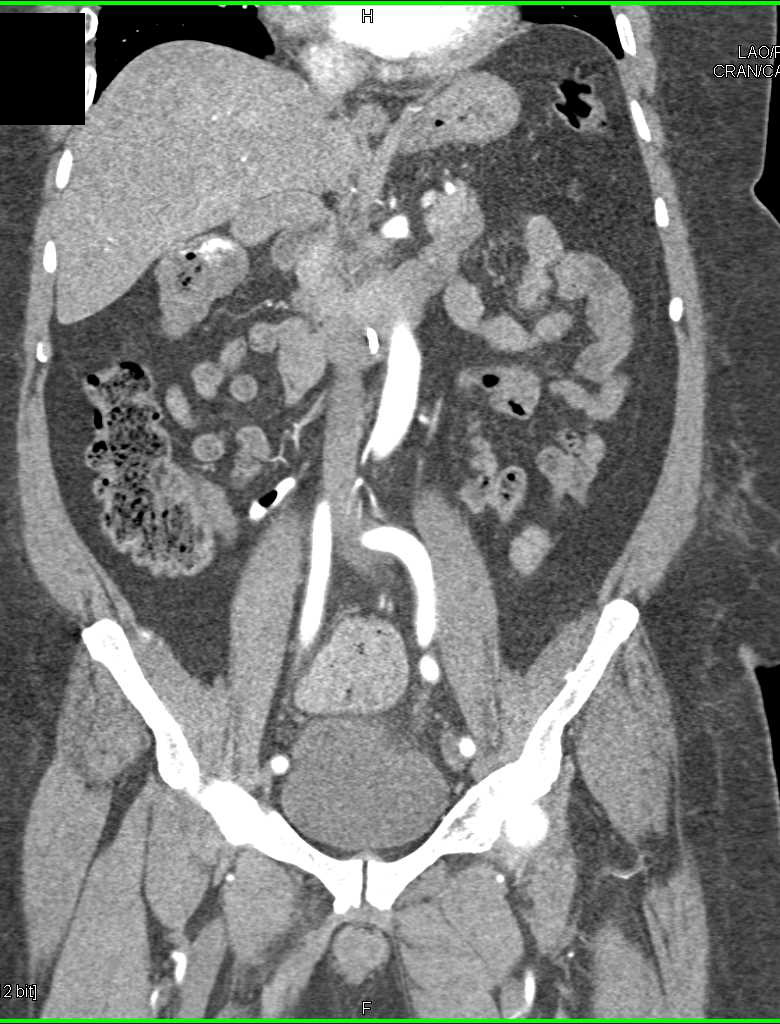
Another way to avoid rib shadows is to ask the patient to inhale. In this case, the probe can be rotated around its axis, with the marker pointing slightly posteriorly (to get in between the ribs). Ribs may get in the way of a clear picture. It may be necessary to move one intercostal space inferiorly to evaluate the liver tip. At this point, the probe is tilted around to assess Morrison’s pouch, liver, and kidney. The hepatorenal recess should be placed in the center of the ultrasound screen. Ideally, the kidney, liver, and diaphragm are seen at the same time. A transducer is put in the mid-axillary line at the lever of the 10 th rib, with the marker on the probe pointing towards the patient’s head. The liver, right kidney, and Morrison’s pouch are assessed using ultrasound in the perihepatic window. The segmental anatomy system is important for surgical procedures, for many pathologies it is however sufficeint to talk abot the left or right lobes of the liver. The liver is divided into two halfs and aditionally in several segments.

The ligamentum venosum separates the caudate lobe from the rest of the liver. The ligamentum teres descends towards the inferoanterior aspect of the liver. At the superior margin of the liver, there is the falciform ligament, splitting the left and right lobes. The hyperechoic, linear structures on liver ultrasounds are the ligaments. The hepatic veins do not have a fibrous sheath and their walls are therefore less reflective than the walls of the portal venous system The three main hepatic veins, left, middle and right, can be traced into the inferior vena cava (IVC) at the superior margin of the liver. However the larger, proximal branches can be demonstrated. In the peripheral parts of the liver the hepatic artery branches and biliary ducts are too small to be detected by ultrasound. The There are blood vessels that are seen as branching tubular structures that can be traced towards the portal vein or the inferior vena cava. A thin, hyperechoic capsule surrounds the smooth parenchyma, which is interrupted by ligaments and vessels. A complete examination of the liver requires scanning from multiple angles and directions.Ī healthy liver ultrasound shows a homogenous, sponge like texture of mid-grey entity, with the same or slightly increased echogenicity as the cortex of the right kidney. The liver is a large organ and therefore cannot be scanned adequately from one approach. The liver offers an outstanding acoustic window on to other organs and vessels in the upper part of abdomen. A careful subcostal compression of the abdominal wall using the ultrasound transducer also improves image representation characteristics through the displacement of the superimposed intestinal structures as well as optimization of the beam angle and distance to the organ. This facilitates access to the ultrasound picture of the liver.

Caudal repositioning can be achieved by asking a patient to take a deep abdominal breath. The liver lies primarily in a high subcostal position. It lies in contact with the right kidney, right adrenal gland, right colic flexure, transverse colon, the first part of the duodenum, gallbladder, esophagus, and the stomach. The visceral surface is posteroinferior and covered with peritoneum, except for the fossa of the gallbladder and porta hepatis. The diaphragmatic surface is anterosuperior, smooth and convex, fitting beneath the curvature of the diaphragm. There are two liver surfaces: the diaphragmatic and visceral surface. The lesser omentum attaches the liver to the lesser curvature of the stomach and first part of the duodenum. The r ight triangular ligament attaches the right lobe of the liver to the diaphragm. The left triangular ligament attaches the left lobe of the liver to the diaphragm. The coronary ligament (anterior and posterior folds) attaches the superior surface of the liver to the inferior surface of the diaphragm and separates the bare area of the liver.
#Hepatic flexture ultrasound free#
The free edge of this ligament contains the round ligament of the liver ( ligamentum teres), a remnant of the umbilical vein. The falciform ligament is the sickle-shaped ligament that connects the anterior surface of the liver to the anterior abdominal wall and forms a natural anatomical division between the left and right lobes of the liver.

They attach the liver to the surrounding structures. There are various ligaments formed by a double layer of peritoneum.


 0 kommentar(er)
